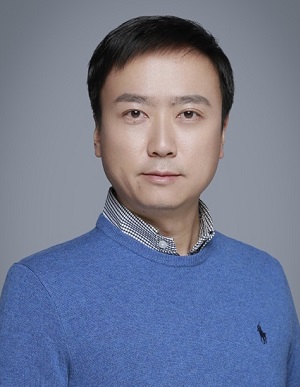

国家"杰出青年科学基金"获得者
中国科学院生物物理研究所,生物大分子全国重点实验室,研究组长
2002 - 2006 浙江大学,光电信息工程学系,学士
2006 - 2011 香港科技大学,电子与计算机工程学系,博士
2011 - 2015 霍华德·休斯医学研究所,Janelia研究校园,博士后
2015 -2024.6 中国科学院生物物理研究所,研究员
2021年 中国科学院优秀青年科学家奖
2021年 国家杰出青年科学基金
2020年 中国科学院优秀导师奖
2019年 掠入射结构光超分辨成像技术发展与应用(Cell, 2018)入选年度中国十大科学进展
2019年 科学探索奖,腾讯基金会
2019年 中源协和生命医学创新突破奖
2015年 PicoQuant 青年研究员奖,国际光学工程学会(SPIE)
2011年 青年科学家奖获奖者,香港科学会
光学显微成像技术已成为生命科学研究中必不可少的共性关键技术,其革新一直是生物医学领域发展的重要驱动力。近年来以超分辨显微镜,单分子成像与追踪,以及多光子非线性显微成像为代表的技术革新,为生命科学发展带来了新的契机。李栋课题组致力于光学显微成像技术的研制工作,先后开发了:(1)条纹激活非线性结构光显微镜; (2)掠入射结构光超分辨显微镜;(3)傅立叶域注意力深度学习超分辨成像等技术方法,并利用这些新型成像技术开展细胞生物学研究。
1. 多模态、高速、多色、长时程超分辨显微镜系统研制
超分辨成像技术为提高分辨率往往需要很高的激发光功率,较长的采集时间,这导致活体样品的漂白和损伤,以及在超分辨图像中产生伪信息。李栋课题组探索新的超分辨成像技术方案,降低获得高可信度超分辨信息的代价,并在获得更高分辨率的同时提高成像速度,延长超分辨成像时程,使其适用更广的生物医学研究领域。
2. 深度学习超分辨显微成像算法开发
生物显微成像技术的发展需要多学科的交叉融合。近年来基于神经网络的深度学习算法有了重大突破,但是将深度学习等人工智能方法用于显微成像技术领域尚处于起步阶段,首先严重缺乏足够丰富且多样的生物图像训练数据,其次面对复杂多样的生物结构,以及不同信噪比和分辨率的成像条件,生物学家在多大程度上能够信任神经网络输出的超分辨图像?如何根据显微成像的技术特点优化设计神经网络?李栋课题组围绕上述问题,结合不同显微成像方法的技术原理,发展深度学习超分辨显微成像算法,并进一步开发软硬件融合的智能显微镜系统。
3. 新型超分辨成像技术应用于生命科学研究
李栋课题组已研制成功的新型显微成像技术为在高时空分辨率下连续捕捉细胞内生物过程的动态分子事件提供了切实可用的解决方案。李栋课题组主要以合作研究形式开展多领域生命科学研究,利用高时空分辨成像提供与测序、生化、质谱等方法相互补的时空动态信息。
1. Qiao C#, Li D#, Liu Y#, Zhang S#, Liu K, Liu C, Guo Y, Jiang T, Fang C, Li N, Zeng Y, He K, Zhu X, Lippincott-Schwartz J*, Dai Q*, Li D*. Rationalized deep learning super-resolution microscopy for sustained live imaging of rapid subcellular processes. Nature Biotechnology, Published: 06 Oct 2022.
2. Andrews B, Chang J, Collinson L, Li D, Lundberg E, Mahamid J, Manley S, Mhlanga M, Nakano A, Sch?neberg J, Valen D, Wu T, Zaritsky A. Imaging cell biology. Nature Cell Biology, 2022 Aug; 24(8):1180-1185. (invited Viewpoint paper)
3. Jiang A#, Zhang S#, Wang X*, Li D*. RBM15 modulates STYK1 m6A to promote tumorigenesis. Comput Struct Biotechnol J, 2022 Sep 5;20:4825-4836.
4. Gong B#, Guo Y#, Ding S, Liu X, Meng A, Li D*, Jia S*, A Golgi-derived vesicle potentiates PtdIns4P to PtdIns3P conversion for endosome fission. Nature Cell Biology, 2021 Jul; 23(7):782-795.
5. Xu G#, Liu C#, Zhou S#, Li Q, Feng Y, Sun P, Feng H, Gao Y, Zhu J, Luo X, Zhan Q, Liu S, Zhu S, Deng H*, Li D*, Gao P*. Viral tegument proteins restrict cGAS-DNA phase separation to mediate immune evasion. Molecular Cell, 2021 Jul 1;81(13):2823-2837.
6. Li D#, Colin-York H#, Barbieri L#, Javanmardi Y#, Guo Y, Korobchevskaya K, Moeendarbary E*, Li D*, Fritzsche M*. Astigmatic traction force microscopy (aTFM). Nature Communications, 2021 Apr 12; 12(1):2168.
7. Barbieri L#, Colin-York H#, Korobchevskaya K#, Li D, Wolfson DL, Karedla N, Schneider F, Ahluwalia BS, Seternes T, Dalmo RA, Dustin ML, Li D*, Fritzsche M*. Two-dimensional TIRF-SIM-traction force microscopy (2D TIRF-SIM-TFM). Nature Communications, 2021 Apr 12;12(1):2169.
8. Qiao C#, Chen X#, Zhang S#, Li D, Guo Y, Dai Q*, Li D*. 3D Structured Illumination Microscopy via Channel Attention Generative Adversarial Network. IEEE J SEL TOP QUANT. 2021 Feb 22;27(4):1-11.
9. Qiao C#, Li D#, Guo Y#, Liu C#, Jiang T, Dai Q*, Li D*. Evaluation and development of deep neural networks for image super-resolution in optical microscopy. Nature Methods, 2021 Feb;18(2):194-202.
10. Wang X#*, Liu C#, Zhang S#, Yan H, Zhang L, Jiang A, Liu Y, Feng Y, Li D, Guo Y, Hu X, Lin Y, Bu P, Li D*. N6-methyladenosine modification of MALAT1 promotes metastasis via reshaping nuclear speckles. Developmental Cell, 2021 Mar 8;56(5):702-715.
11. Qin J, Guo Y, Xue B, Shi P, Chen Y, Su Q, Hao H, Zhao S, Wu C, Yu L, Li D*, Sun Y*. ER-mitochondria contacts promote mtDNA nucleoids active transportation via mitochondrial dynamic tubulation. Nature Communications, 2020 Sep 8;11(1):4471.
12. Li D#, Zhao Y#, Li D, Zhao H, Huang J, Miao G, Feng D, Liu P, Li D*, Zhang H *. The ER-Localized Protein DFCP1 Modulates ER-Lipid Droplet Contact Formation. Cell Reports, 2019 Apr 9;27(2):343-358.
13. Colin-York H#, Javanmardi Y#, Barbieri L, Li D, Korobchevskaya K, Guo Y, Hall C, Taylor A, Khuon S, Sheridan G, Chew T, Li D*, Moeendarbary E*, Fritzsche M*. Spatiotemporally Super-Resolved Volumetric Traction Force Microscopy. Nano Letters, 2019 Jul 10;19(7):4427-4434.
14. Guo Y #, Li D#, Zhang S, Yang Y, Liu JJ, Wang X, Liu C, Milkie DE, Moore RP, Tulu US, Kiehart DP, Hu J, Lippincott-Schwartz J*, Betzig E*, Li D*. Visualizing intracellular organelle and cytoskeletal interactions at nanoscale resolution on millisecond timescales. Cell, 2018 Nov 15;175(5):1430-1442.
15. Han X #, Li P #, Yang Z, Huang X, Wei G, Sun Y, Kang X, Hu X, Deng Q, Chen L, He A, Huo Y, Li D*, Betzig E, Luo J*. Zyxin regulates endothelial von Willebrand factor secretion by reorganizing actin filaments around exocytic granules. Nature Communications, 2017 Mar 3; 8:14639.
16. Li D, Shao L, Chen BC, Zhang X, Zhang M, Moses B, Milkie DE, Beach JR, Hammer JA, Pasham M, Kirchhausen T, Baird MA, Davidson MW, Xu P, Betzig E*. Extended-resolution structured illumination imaging of endocytic and cytoskeletal dynamics. Science, 2015 Aug 28;349(6251): aab3500. Cover article.
17. Beach JR*, Shao L, Remmert K, Li D, Betzig E, Hammer JA*. Nonmuscle myosin II isoforms coassemble in living cells. Current Biology, 2014 May 19;24(10):1160-1166.
18. Li D#, Zheng W#, Zeng Y, Luo Y, Qu JY*. Two-photon excited hemoglobin fluorescence provides contrast mechanism for label-free imaging of microvasculature in vivo. Optics Letters, 2011 Mar 15;36(6):834-836.
19. Li D#, heng W#, Zhang W, Teh SK, Zeng Y, Luo Y, Qu JY*. Time-resolved detection enables standard two-photon fluorescence microscope for in vivo label-free imaging of microvasculature in tissue. Optics Letters, 2011 Jul 15;36(14):2638-2640.
20. Li D, Zheng W, Zeng Y, Qu JY*. In vivo and simultaneous multimodal imaging: integrated multiplex coherent anti-Stokes Raman scattering and two-photon microscopy. Applied Physics Letters, 2010 97(22): 223702.
21. Li D, Zheng W, Qu JY*. Imaging of epithelial tissue in vivo based on excitation of multiple endogenous nonlinear optical signals. Optics Letters, 2009 Sep 15;34(18):2853-2855.
22. Li D, Zheng W, Qu JY*. Two-photon autofluorescence microscopy of multicolor excitation. Optics Letters, 2009 Jan 15;34(2):202-204.
23. Li D, Zheng W, Qu JY*. Time-resolved spectroscopic imaging reveals the fundamentals of cellular NADH fluorescence. Optics Letters, 2008 Oct 15;33(20):2365-2367.
(资料来源:李栋研究员,2023-01-12)